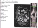
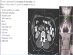
بخشی از پاورپوینت
--- پاورپوینت شامل تصاویر میباشد ----
اسلاید 1 :
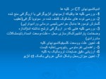
اندیکاسیونهای CT در کلیه ها
1- بررسی کلیه ها وقتیکه آزمونهای آنژیوگرافی یا اروگرافی منع شده
2- بررسی توده های مشکوک کشف شده در سونویا گرافی(تعقیب گسترش تومورها بدنبال جراحی,شیمی درمانی ورادیوتراپی)
3- بررسی کلیه هایی که در اروگرافی ترشح نداشته اند(اندازه وضخامت پارانشیم,آشکارسازی محل –علت-وسعت انسداد,تایدمشکلات مادرزادی)
4- تعیین علت ووسعت کلسیفیکاسیونهای کلیه
5- راهنمایی نفرستومی ,بایوپسی,تخلیه کیست
6- ارزیابی دقیق ضایعات تروماتیک به کلیه
7- تعیین میزان,محل وشکل تنگی عروقی بکمک ct آنژیو
اسلاید 2 :
-در اسکن بدون کنتراست پارانشیم طبیعی کلیه عدد ctحدود 50-30 داشته وبکمک تزریق سریع کنتراست در اسپیرال کورتکس از مدولا بخوبی تفکیک می شود.
- C t کلیه باید با کنتراست خوراکی بمنظور افتراق لوپهای روده از توده های ناحیه وآدنوپاتی خلف صفاقی انجام گیرد.
-اسکن با دو فاز با وبدون کنتراست انجام می شود.اسکن بدون کنتراست جهت بررسی کلسیفیکاسیونهای پارانشیمال, وتومورهای چربی مفید است . سنگهای کوچک ادراری,خونریزیهای وچربیهای دورکلیوی قبل از محو شدن توسط کنتراست انجام می شود.تشخیص افتراقی کیستهای هایپردنس از تومورهای کلیوی. CTkuB اختصاصا در ارزیابی سنگها ی ادراری حاد بدون هرگونه کنتراست از قطب فوقانی کلیه تاسمفیز با برشهای 3 پیچ 5/1انجام می شود.
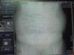
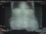
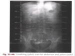
-توپوگرام از بالای کلیه ها تا کف مثانه AP
-
اسلاید 3 :
Aquisition Technique
Depending on the clinical imaging task, the required spatial resolution varies. For most dedicated indications for the kidneys as well as for obstructive uropathy, a section collimation of 3-5 mm should be used. The smaller the collimation,the better the quality of multi planar reformations of the renal parenchyma and excretory system. In addition, the differentiation of small cysts from tumors, as well as the detection of calculi, improves because of less partial volume effects With multislice CT, moderately thin sections (4 x 2.5 mm) suffice for most indications although the quality of coronal reformats is substantially improved with 0.5-1.25 mm collimation on 4- to 16-slice scanners. Such thin-section protocols become important, however, for a subtle evaluation of the renal calyces.
اسلاید 4 :
For obstructive uropathy, best image quality is obtained
with a thin-section, 0.5-1.25 mm protocol and curved planar reformats(CPR) along the course of the ureter . A 4 x 2.5 mm protocol will allow for a somewhat lower patient dose (only relevant in 4-slice scanners ).Coronal reformats should become standard for optimum demonstration of the extent and location of lesions. Unlike MRI, the coronal
plane can be optimally adjusted after the data set has been acquired. Image quality often can be improved if thick MPR(section width 3 mm) are employed. Maximum intensity projections may be helpful to demonstrate renal calculi and
to demonstrate the urinary tract during the excretory
phase.
اسلاید 5 :
Usually a 3 -5 mm slice with a pitch of 1 (table speed 5 mm/sec) and reconstruction every 3mm is used. However if the patient is large, weighing more than 100 kg, a 5 mm slice should be used as there is a limit to the mA value that is available and the images may be very noisy.
- Scan delay depends on whether corticomedullary images (50 second delay) or nephrogram images (100-180 second delay) arerequired.
اسلاید 6 :
SCOUT: AP
LANDMARK: XIPHOID TIP
SCANNING MODE: SPIRAL
I.V. CONTRAST: 2-4 ml/sec
SCAN DELAY:1. NONCONTRAST: 2. ARTERIAL 30 SEC.
3. NEPHROGRAM 90 SEC.:
4. PYELOGRAM 3-5 MIN.
ORAL CONTRAST: 400 ml 45 MINUTES BEFORE SCAN,
200 ml JUST BEFORE SCAN
BREATH HOLD: SUSPENDED EXPIRATION
SLICE THICKNESS: 3-5 MM , 5 MM THROUGH
KIDNEYS
START LOCATION: LUNG BASES
END LOCATION: ILIAC CREST
FILMING: STANDARD
اسلاید 7 :
:Patient position
Supine with elevated arms
Windowing:
Noncontrast CT: W/L=300/40
Contrast-enhanced CT: W/L=400-500/50-100
FILMING:
STANDARD
اسلاید 8 :
فاز كورتيكومدولاري اين فاز وقتي آغاز مي شود كه مواد كنتراست وارد مويرگ هاي قشري و فضاي peritubular شده و به توبول هاي كورتيكال پروگزيمال فيلتر ميشود.
فاز كورتيكو مدولاري بين 25 تا 70 ثانيه بعداز شروع تزريق پديدار مي شود.
Scanning during the corticomedullary phase
(arterial phase, vascular phase, vascular nephrogram
phase, delay 20-35 seconds) is a useful
addition for demonstrating the vascular anat-
0my of the kidneys, especially in patients
scheduled for nephron-sparing surgery.
اسلاید 9 :
فاز نفروگرافيک
Parenchymalphase
اين فاز وقتي آغاز مي شود كه مواد حاجب از عروق كورتيكال و فضاي بينابيني خارج سلولي گذشته و به قوس هاي هنله و توبول هاي جمع كننده وارد مي شود.
اين فاز حدود 80 ثانيه بعد ازشروع تزريق آغازشده وحداكثر 180ثانيه بعداز تزریق ادامه دارد.
اين فاز براي آشكار نمودن توده هاي كليوي وشناخت و تشخيص ضايعات نامعين بسيار با ارزش است.
اسلاید 10 :
فاز ترشحي
اين فاز تقريبا 3-5دقیقه بعد از شروع تزريق آغاز ميشود.
ماده حاجب به داخل سيستم جمع آوري كننده ترشح شده ودانسيته فاز نفروگرام به ميزان قابل توجهي كاهش مي يابد.
اين فاز براي بررسي توده هاي يوروتليال كمك كننده است.

